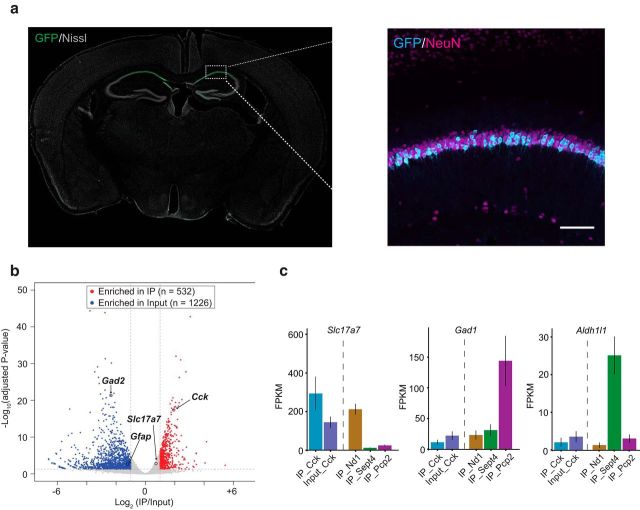
Figure 2.
Characterization of Cck-bacTRAP line and immunoprecipitation of ribosome associated mRNAs from hippocampal CA1 neurons. a, Coronal section of a Cck-bacTRAP mouse brain stained for GFP and Nissl. Inset picture is an immunohistochemistry image of hippocampus with antibodies against GFP (cyan) and NeuN (magenta) showing colocalization in CA1. Scale bar, 100 μm. b, Volcano plot analysis revealed transcripts specifically enriched in Cck-bacTRAP IP samples versus input samples (whole hippocampus lysates). Red dots represent transcripts that were significantly enriched in IP samples (e.g., Cck) and blue dots represent transcripts that were significantly enriched in input samples (e.g., Gfap, Gad2). c, Quantification of expression levels of Slc17a7 (Vglut1, excitatory neuron marker), Gad1 (inhibitory neuron marker), and Aldh1l1 (glial marker) across different bacTRAP lines expressing in different cell types: Cck (hippocampal CA1, from current study), Nd1 (cerebellar granule neuron), Sept4 (cerebellar Bergmann glia), Pcp2 (cerebellar Purkinje cell); the latter three are all published datasets from Dr. Heintz's group (Mellén et al., 2012). FPKM, Fragments per kilobase of transcript per million mapped reads.

