
Figure 3. Artery and vein specification in the developing pancreas.
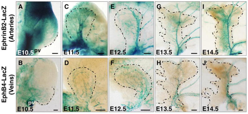
(A, C, E, G, I) EphrinB2-lacZ pancreata at indicated stages stained for β-Galactosidase activity in whole mount to label arteries. (B, D, F, H, J) EphB4-lacZ pancreata at indicated stages stained for β-Galactosidase activity in whole mount to mark veins. Dashed line represents the boundary of pancreatic bud. Scale bars 100 μm. pv, portal vein.