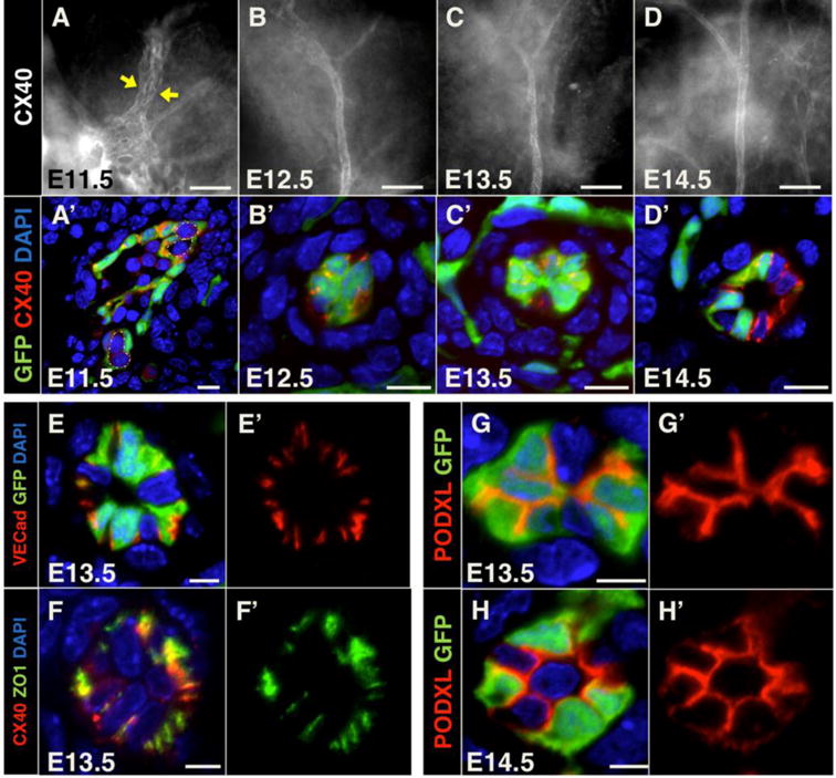
Figure 5. Development of the central artery in the pancreas via vessel coalescence.

(A, B, C, D) Whole mount pancreata at indicated stages immunostained for the artery marker Connexin40 (CX40). Yellow arrows indicate coalescing vessels marked by Connexin40. Scale bars 50 μm. (A′, B′, C′, D′) Cryosections of Flk1-GFP pancreata at indicated stages immunostained for GFP (green) and Connexin40 (red) to mark endothelial cells and arteries, respectively. Dashed lines in A′ represent lumen boundaries. Scale bars 10 μm. (E–E′) Cryosections of E13.5 Flk1-GFP pancreata immunostained for GFP (green) and the endothelial junctional marker VE-cadherin (red). Scale bars 5 μm. (F–F′) Cryosections of E13.5 pancreata immunostained for the tight junction molecule Zona Occludens 1 (ZO1, green) and Connexin40 (red). Scale bars 5 μm. (G–H′) Cryosections of E13.5 and E14.5 Flk1-GFP pancreata immunostained for GFP (green) and the apical glycoprotein PODXL (red). Scale bars 5 μm. DAPI (blue) marks nuclei when applicable.
