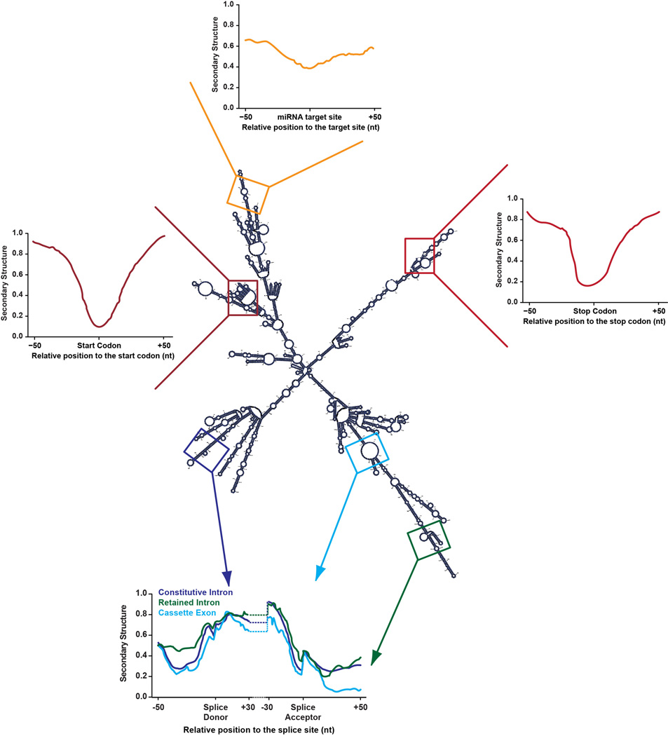
Figure 2. Structural patterns in mRNAs.
This is an example of a folded Arabidopsis thaliana mRNA. The displayed secondary structure profiles are representative of metagene patterns at start codons (dark red) miRNA target sites (orange), stop codons (red), constitutive intron (purple), retained intron (green), and cassette exon (blue) splice donor sites.

