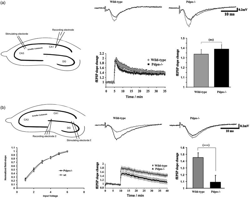
Figure 2.
Podoplanin deletion selectively impairs long-term synaptic plasticity in the hippocampal dentate gyrus. (a) Comparative examinations of LTP in CA3-Schaffer collateral-CA1 synapses (inset cartoon in left represents the positioning of stimulating and recording electrodes) of slices obtained from wild-type and podoplanin−/− mice (n = 9 animals per group). (b) No differences in basal synaptic transmission (left) was detected at the dentate gyrus (inset cartoon in left represents the positioning of stimulating and recording electrodes) in hippocampal slices from podoplanin−/− and control wild-type mice as examined by input/output protocols. Right, representative fEPSPs traces (upper insets); temporal courses (lower chart plot) of averaged field-postsynaptic-potential-slopes; and corresponding bar graphs of the end-time points (right) of data obtained before and after application of electrical stimulation inducing long-term-potentiation (LTP) in the dentate gyrus. A mixed-model repeated measure ANOVA indicated no main effect of genotype (p > 0.05), a highly significant main effect of time (F158,82 = 57.05, p < 0.0001) and a significant interaction between the two factors (F52,16 = 3.45, p < 0.0001). Data revealed significant differences in the later phases of synaptic potentiation only in the dentate gyrus. Data are displayed as mean ± SEM. ***(p ≤ 0.001), ns (p > 0.05).

