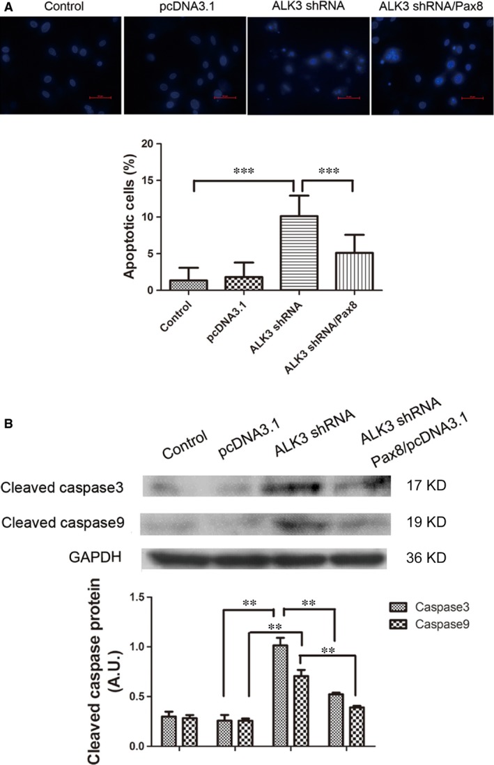
Figure 3.

Pax8 rescued apoptosis induced by ALK3 silence in H9C2 cells. (A) Apoptosis was observed by DAPI staining by using a fluorescence microscopy. Positive apoptotic cells were indicated by bright blue staining. Ten different visual fields were randomly chosen to count positive cells. The percentage of positive cells relative to the total cells was considered as the rate of apoptosis in each visual field. (B) The H9C2 cells performed as a control group, negative control were transfected with pcDNA3.1and pGPU6 vectors. The ALK3 silence group was transfected with ALK3 shRNA/pGPU6. Treating group was pre‐treated with Pax8/pcDNA3.1 24 hrs earlier and ALK3 shRNA/pGPU6. Data are presented as mean ± S.D. **, P < 0.01. ***, P < 0.001 (magnification: ×400).
