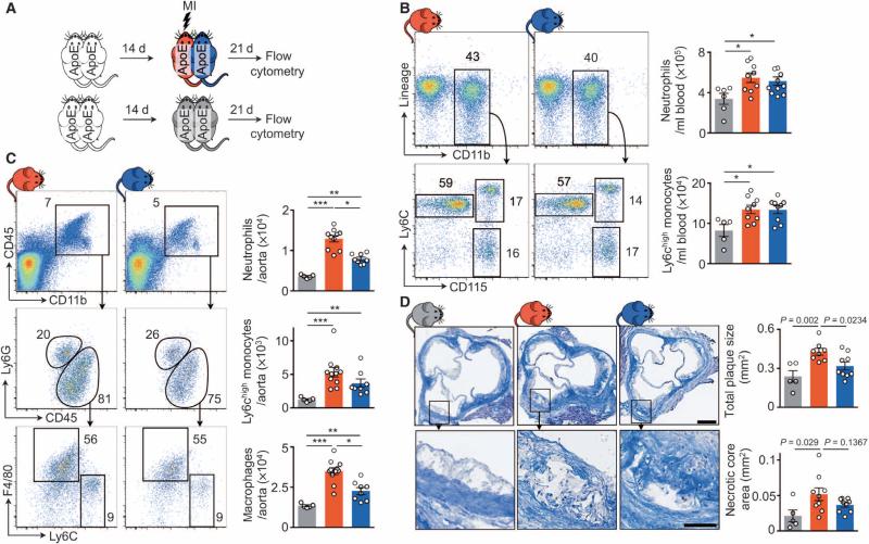
Fig. 1. Noncirculating factors contribute to expansion of inflammatory plaque leukocytes and acceleration of atherosclerosis.
(A) Experimental setup. ApoE−/− mice were joined in parabiosis, and one parabiont was infarcted 2 weeks thereafter. Three weeks later, leukocytes were evaluated by flow cytometry. (B and C) Flow cytometric gating and quantification of blood (B) and aortic (C) leukocytes. Data are means ± SEM (n = 5 to 11 per group from two independent experiments). *P < 0.05, **P < 0.01, ***P < 0.001, two-tailed paired Wilcoxon test for comparison between connected parabionts; two-tailed unpaired Mann-Whitney U for comparison to steady-state parabiosis. (D) Masson staining of aortic roots. Data are means ± SEM quantifying total plaque size and necrotic core area per section (n = 5 to 9 per group from two independent experiments). Scale bars, 250 μm (top) and 100 μm (bottom).

