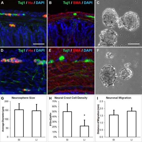
FIGURE 1. Neurospheres can be generated from the small and large intestine of mice.
Enteric ganglia containing Tuj1+ and Hu+ neurons are found in both small intestine (SI, A) and large intestine (LI, D). Tuj1+ neurons in the myenteric plexus were isolated together with the surrounding SMA+ muscularis propria from SI (B) and LI (E) and neurospheres were formed (C, F; respectively). While neurospheres from both sources were similar in size (G), SI-derived neurospheres contained significantly more P75+ neural crest-derived cells (H). Migratory distance of neurons from SI- and LI-derived neurospheres was similar (I). Scale bar in A is 100 μm and applies to A-B and D-E. Scale bar in C is 100 μm and applies to C and F.
*p<0.05; SMA, smooth muscle actin; SI, small intestine; LI, large intestine

