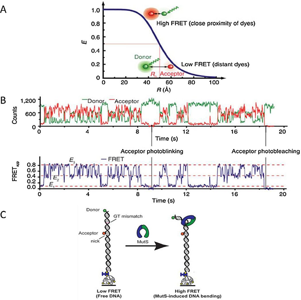
Fig. 4.
Förster resonance energy transfer. (A) During FRET a donor dye is excited and in a non-radiative process this excitation energy is transferred to an acceptor dye. The FRET efficiency (E) of this process depends upon distance between dyes (R), accurate measurement of distance is made around the mid-point of FRET efficiency – the Förster distance. (B) An example of two color single molecule FRET data. The intensities of the individual donor and acceptor dyes (upper trace) permit calculation of the FRET efficiency (lower trace) to be calculated. Reprinted (adapted) with permission from [37]. Copyright (2013) American Chemical Society. (C) smFRET system with MutS-induced DNA bending. A 50 base pair strand of dsDNA with a FRET donor (green) and a FRET acceptor (red) is anchored to a surface through a biotin streptavidin link. A GT-mismatch is located approximately halfway between the dyes. As the MutS binds to the DNA and bends the DNA the distance between the dyes decreases which increases the FRET signal.
Reprinted (adapted) with permission from [39]. Copyright (2013) American Chemical Society.

