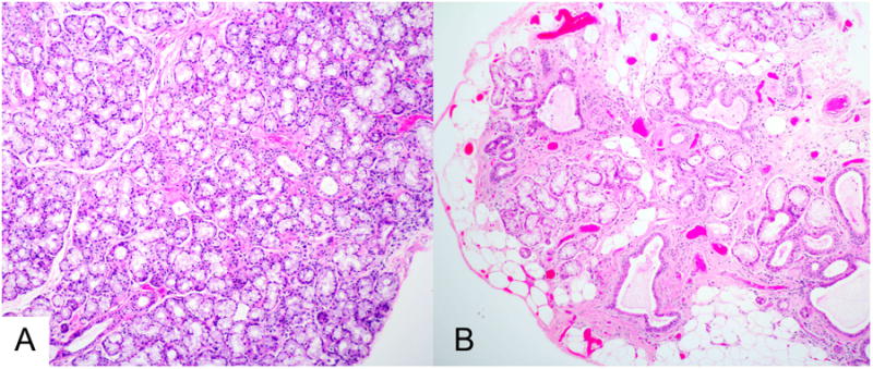
Figure 1.

Histopathology of minor labial salivary glands. The sections are from biopsies of a 28 year-old woman (panel A) and a 65 year-old woman (panel B), shown at the same magnification. The histopathologic section in panel A shows normal tissue, with confluent mucous acini and normal-sized intralobular ducts. In contrast, the section in panel B shows extensive acinar loss, interstitial fibrosis, ductal dilatation, and fatty replacement. The changes in panel B are often seen to varying degree in older patients. (Magnification 100 ×)
