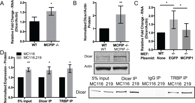
Fig 6. Repression of MCPIP1 increases Dicer expression and TRBP association.
(A) RNA from WT and MCPIP1 -/- MEFs were analyzed for Dicer expression using qPCR. Results are shown relative to β-actin and normalized to the WT control. N = 3. (B) WT and MCPIP1 -/- MEF protein lysates were evaluated for Dicer using western blot analysis. β-actin was used as a loading control. The results are averaged for six replicates, and a representative western blot is shown. (C) WT and MCPIP1 -/- MEFs were transfected with a MCPIP1 expression vector or an EGFP negative control vector, and RNA was isolated 48 hpt. RNA was DNase treated and analyzed for Dicer expression using qPCR. Results are shown relative to β-actin and normalized to the WT control, N = 4. (D) MC116 and KSHV infected MC116.219 cells were immunoprecipitated with anti-Dicer and anti-TRBP antibodies and blotted for Dicer. The results are averaged for four replicates and a representative blot is shown. For all graphs, results are shown as mean ± SD. Significance was assessed using a Student’s t test, *p ≤ 0.05, **p ≤ 0.01. Numerical data can be found in S1 Data.

