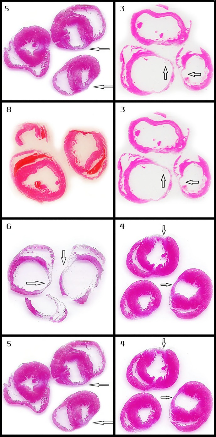
Fig 5. Comparison of myocardial axial section of different protocols.
Every row in the figure compares the axial section of the left ventricle of rats that were treated with two different IRE protocols. Moreover, all the pairs of protocols selected for this figure are different in only one setting of the IRE protocols (i.e. voltage, frequency, etc.) and demonstrate significant differences between their morphometric measurements. Protocol numbers are in the right upper corner of each slide. The pairs of protocols are (from top to bottom): 5 and 3(70μsec and 100μsec), 8 and 3 (MI vs. IRE), 6 and 4 (10 pulses vs. 20 pulses), 5 and 4 (2Hz vs. 1HZ).

