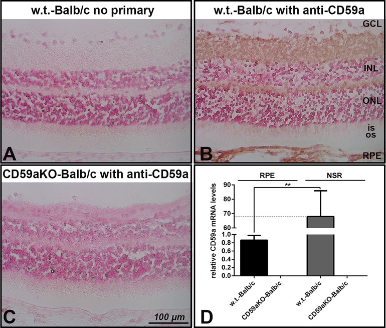
Fig 1. CD59a expression in mouse retina and mRNA levels of CD59a in NSR and isolated RPE.
Photomicrograph showing CD59a immunolabeling in all layers of mouse retina, RPE and choroid (B), but not in CD59aKO eyes (C). Graph of qPCR results showing that mRNA levels of CD59a in NSR were more than 60-fold higher than that in isolated RPE cells in WT eyes (**P<0.001) (D). Data are expressed as means ± SD. N = 4. RPE, retinal pigment epithelium; OS, photoreceptor outer segment; IS, photoreceptor inner segment; ONL, outer nuclear layer; INL, inner nuclear layer; GCL, ganglion cell layer.

