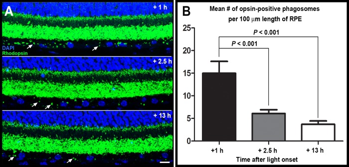
Fig 5. Quantification of RPE phagocytosis of shed outer segment fragments via immunostaining with anti-rhodopsin antibody.
CD59aKO RPE showed maximal phagosome content 1 h after light onset as characteristic for normal outer segment renewal. Representative retinal cross sections (A) showing opsin immunolabeling (green) and cell nuclei (blue) of CD59aKO outer retina of mice sacrificed at 1 h, 2.5 h, and 13 h after light onset as indicated. In each section, two phagosomes in the RPE are indicated by arrows. Scale bar = 10 μm. Quantification of phagosome counts (B). Eyes from 5 different mice for each time point, and 6 sections of each eye were imaged for each mouse. Data are expressed as mean ± SD. N = 5.

