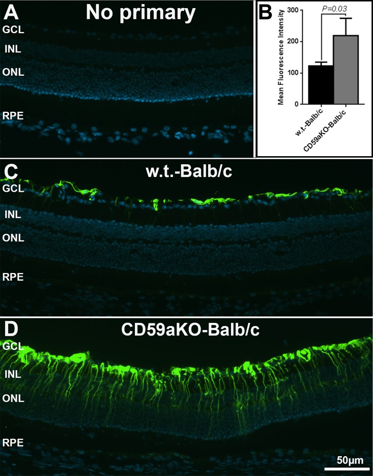
Fig 6. Immunofluorescence microscopy of GFAP in NLD CD59aKO and WT retinas.
Compared to the labeling limited to the innermost retina in WT mice (C), the GFAP signal was up-regulated in Müller cells of CD59aKO retinas (D). Quantification of fluorescence intensity showed that GFAP was significantly increased in CD59aKO retinas compared to controls (B) (P = 0.03). Data are expressed as means ± SD. N = 4.

