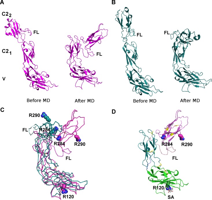
Fig 4. The 3D model for Siglec-9-EC.
The two alternative models for Siglec-9-EC with a differently modeled flexible linker region (FL, between C21 and C22 domains) before and after the refinement by MD simulations. (A) Model 1. (B) Model 2. (C) Superimposition of the V-C21 domains of the two models shows a slightly different orientation for the C22 domain. Model 1 in magenta and model 2 in cyan. (D) The best model for Siglec-9-EC (model 1). Sialic acid (SA; in grey sticks) is modeled to interact with R120 in the V domain. The V domain is shown in green, C21 in blue and C22 in magenta. In (C) and (D), R120 in the V domain and R284 and R290 in the C22 domain are shown as spheres and cysteines forming the disulphide bridges in yellow.

