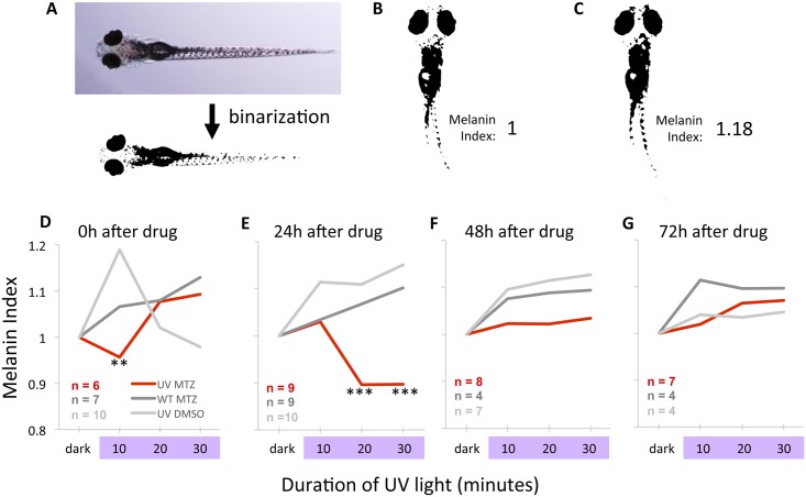
Fig 11. Photoreception of UV light is reduced following ablation of UV cones as measured by changes in body pigmentation.
UV cones were ablated in larvae, and larval body pigmentation was measured in response to UV light; zebrafish larvae of this age are known to darken in response to UV light via melanin dispersion. A. Dorsal view of larval zebrafish. Binarization of images was used to quantify melanosome reaction to UV light. Larvae were imaged at multiple time points, beginning with dark-adapted larvae. B. The number of pixels in dark-adapted larvae was given a melanin index of 1. C. UV light exposure causes wild type fish to darken, increasing the melanin index. D. Shortly after drug treatment (0 hours), control larvae respond to UV light with the expected darkening of body pigmentation (grey lines), whereas larvae with UV cones ablated exhibit a significantly delayed response (red line). E. Larvae tested 24h after drug treatment reveal that UV cone ablation significantly impairs photoreception. F,G. Larvae tested 48 and 72 hours after drug treatment reveal a gradual recovery of sensitivity to UV light to levels indistinguishable from controls. ** = p<0.01 compared to UV+DMSO group *** = p<0.001 comparing UV+MTZ group to larvae in all control groups in the same timepoint, and these data are also significantly different (p<0.01) from the melanin index of the same UV+MTZ larvae in the dark-adapted state. Statistical significance was determined by Two-way ANOVA with a post-hoc Tukey test. The standard error surrounding each mean is reported in Table 3.

