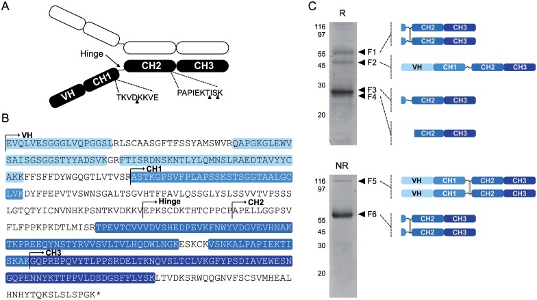
Fig 5. MS/MS characterization of H10 heavy chain fragments isolated from SlCYS8-expressing plants.
A: Schematic diagram for the H10 heavy chain, highlighting the variable VH domain, the constant domains CH1, CH2 and CH3, and the hinge region linking CH1 to CH2. Black arrows point to the most important cleavage sites of H10 heavy chain in the N. benthamiana leaf cell secretory pathway as recently inferred by Hehle et al. [20]. B: Amino acid sequence of H10 heavy chain. Peptide sequences identified by LC-MS/MS (see panel C) are shaded in blue. C. Correspondence between MS/MS unique peptide data and heavy chain domain fragments or assemblies detected on Coomassie blue-stained gels following SDS-PAGE in reducing (R) or non-reducing (NR) conditions. Six fragments (bands F1 to F6) detected following electrophoresis and Coomassie blue staining were excised manually and submitted to LC-MS/MS analysis. Schematic representations on the right indicate the composition of each fragment based on MS/MS data. Sequence details for identified unique peptides are given in S1 Table.

