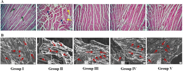
Fig 10.
Histological (Panel A) and ultrastructural (Panel B) assessments of heart of T2D rats of different groups. Group II exhibited degeneration of interstitial tissues (blue arrows) and change in normal radiating pattern (yellow arrows) in the section of heart, while, Group I exhibited general radiating pattern of heart section. SEM showed ventricular portion of araldite sectioned rat myocardial tissues. Myocardial tissue of normal rats (Group I) exhibited normal myocardial fine structure, with myofibrils comprising regular and continuous sarcomeres which demarcated by Z-lines (Red arrow heads), which were in register with adjacent myofibrils and the rows of moderately electron dense mitochondria (Mi) intervene between myofibrils, while, Group II showed randomly distributed mitochondria (Mi) between poorly organized myofibrils in an electron-lucent sarcoplasm. Group III, IV and V indicated significant improvement in myofibrillar arrangement in heart tissues comparable to that of Group I. Group I: Normal control; Group II: T2D control; Group III: T2D rats treated with SR (50 mg/kg, orally); Group IV: T2D rats treated with SR (100 mg/kg, orally); Group V: T2D rats treated with glibenclamide (1 mg/kg, orally).

