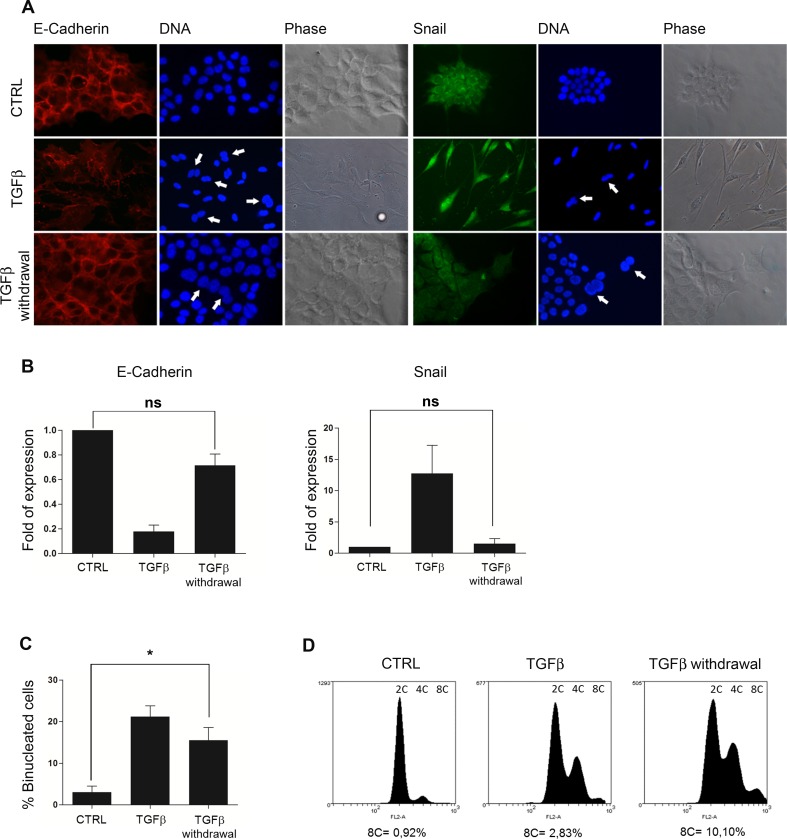
Fig 5. Tetraploid hepatocytes proliferate maintaining the poliploidy state in their progeny after TGFbeta1 withdrawal.
(A) Immunofluorescence for E-Cadherin and Snail of TGFbeta1-treated, TGFbeta1-released and control MMH/E14 hepatocytes. Arrows indicate binucleated cells. (B) Transcriptional analysis by qRT-PCR of E-Cadherin and Snail in the indicated experimental conditions. Data are expressed as average values of three different experiments ± s.e.m. (ns = not significant p value), and plotted as ratio between treated and untreated (CTRL = 1) cells. (C) Percentage of binucleated cells in the indicated experimental conditions. Data are expressed as average values of three different experiments ± s.e.m. (*p value < 0,05). (D) Flow cytometry analysis for DNA content of cells cultured in the indicated experimental conditions.

