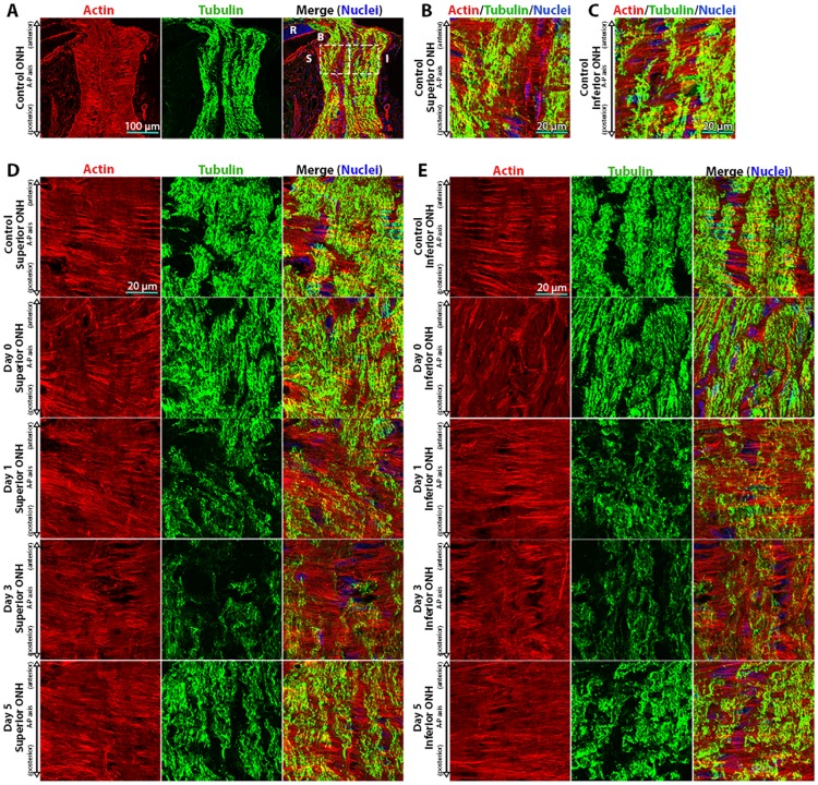
Fig 1. Actin-based astrocyte extensions change orientation within hours after intraocular pressure elevation.
(A) Low magnification images of control optic nerve head (ONH) sections labeled for actin (TRITC-phalloidin), tubulin (Tuj1 anti-tubulin βIII antibody), and nuclei (DAPI). The left and right boxes in the merged image indicate the anterior superior and inferior regions of the ONH, respectively. (B, C) High magnification images of the superior and inferior regions of the ONH, as indicated by boxes in panel (A). Representative superior (D) and inferior (E) ONH regions from control eyes and eyes exposed to 8 hours of intraocular pressure (IOP) elevation, labeled for actin, tubulin, and nuclei. Day 0 eyes were immediately fixed after 8 hours of IOP elevation, while day 1–5 indicate the period of time the IOP was normalized post IOP elevation prior to fixation. A-P = anterior-posterior, B = Bruch’s membrane, I = inferior, R = retina, and S = superior.

