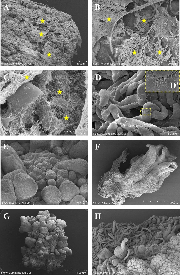
Fig 3. SEM images of the Brachypodium embryogenic callus surface pattern.
A callus on the 7th (A-E) and 21st (F-H) days after cultivation. (A-C)–embryogenic masses covered by an extracellular matrix surface network (ECMSN is indicated by yellow asterisks): note the fibrillar and membranous structure. (D) Parenchymatous cells; high-magnification insert (D’) shows the contact between these cells. (E) Developing somatic embryos. (F) Developed multiple coleoptiles. (G) and (H) a callus on the 21st day after cultivation showing the absence of ECMSN on the surface.

