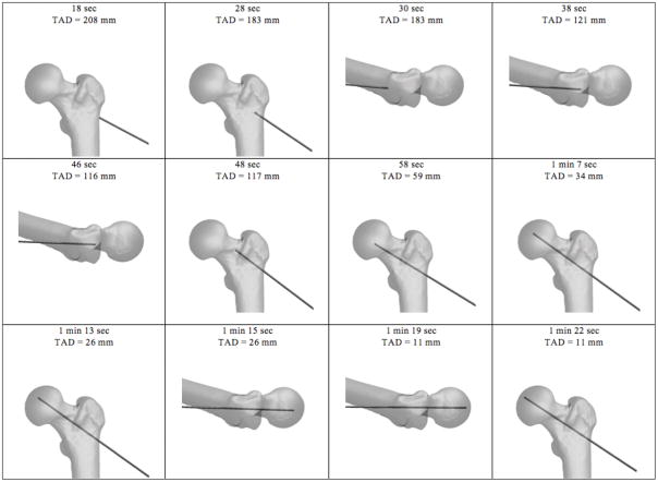
Figure 4.
Computer software utilizes positional data from the electromagnetic sensors to generate AP and lateral fluoroscopic images of the proximal femur and guide pin position. Progress in the simulation is shown going from upper left to right. Time stamp and tip-apex distance measures, which are not shown during the simulation, are included for reference.

