Figure 3.

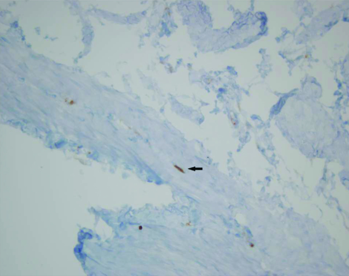
Histopathology of the fetal rat bladder of the control group (arrow indicates interstitial cells of Cajal located in the muscular layer with fusiform cell body, thin cytoplasm, wide ovoid nucleus and two dentritic processes) (C-kit, ×40)

Histopathology of the fetal rat bladder of the control group (arrow indicates interstitial cells of Cajal located in the muscular layer with fusiform cell body, thin cytoplasm, wide ovoid nucleus and two dentritic processes) (C-kit, ×40)