
Figure 1 a–d.
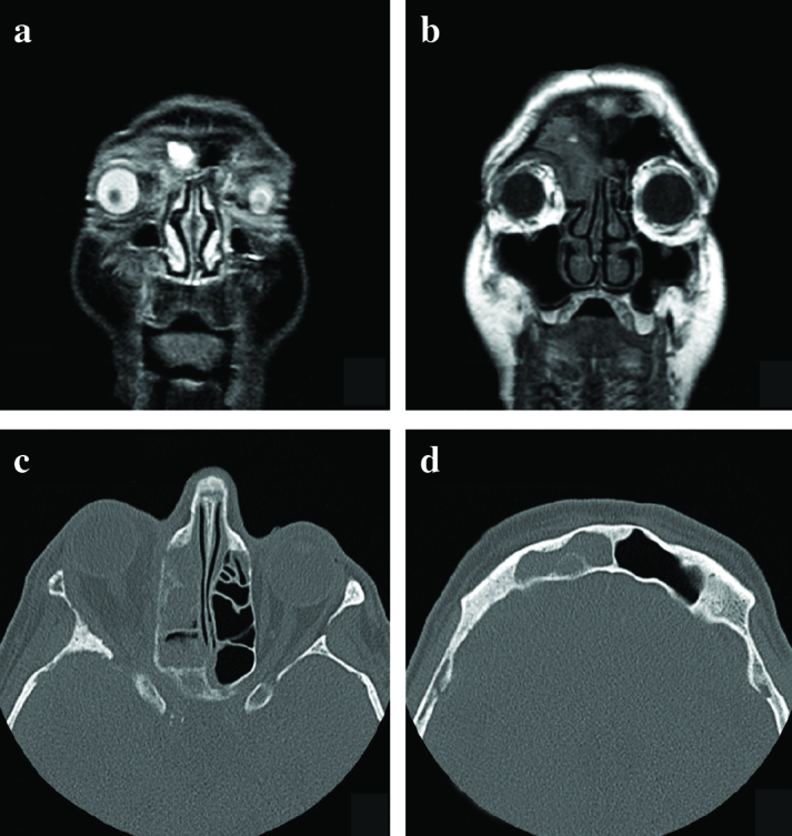
(a) Orbital MRI; Exophthalmos on the right eye and right frontoethmoidal sinus involvement are seen. (b) MR Imaging showing involvement of the frontal, ethmoid sinus. (c, d) Paranasal sinus tomography showing frontoethmoidal sinus involvement