Figure 7.
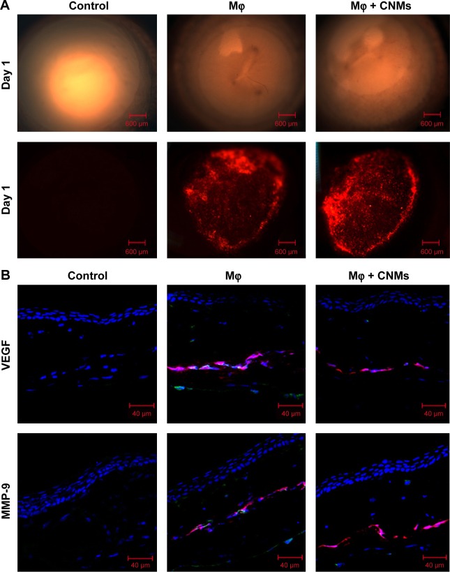
In vivo fluorescence imaging and effect of CNMs on VEGF and MMP-9 expression in rat cornea.
Notes: (A) Fluorescence micrographs of a cornea indicate the Mφ labeled with DiI located in cornea pockets on postoperative day 1 in the Mφ group and CNMs group. There were no DiI-labeled cells in the cornea pocket in the control group. Magnification ×25. (B) Confocal laser scanning microscopic images of a cornea indicate the protein expression of VEGF and MMP-9 on postoperative day 2. Visibly high expression of VEGF and MMP-9 was observed in implanted DiI-labeled Mφ, corneal stroma, and corneal endothelium in the Mφ group. There was a significant reduction in VEGF and MMP-9 expression in implanted Mφ and corneal stroma and endothelium in the CNMs group. No remarkable expression of VEGF and MMP-9 was observed in the control group.
Abbreviations: CNMs, celastrol nanomicelles; DiI, 1,19-dioctadecyl-3-3-39,39-tetramethylindocarbocyanine; Mφ, macrophage; MMP-9, matrix metalloproteinases 9; VEGF, vascular endothelial growth factor.

