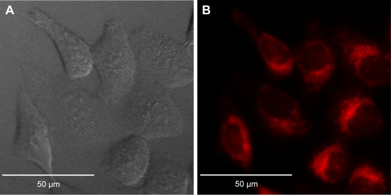
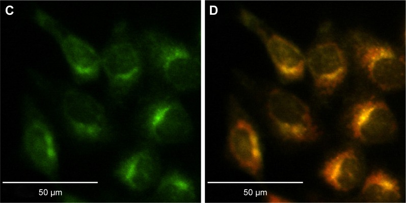
Figure 4.
Lysosome colocalization experiments.
Notes: (A) DIC and confocal fluorescence micrographs of HCT-116 cells incubated with PEG–PAG9 (B; red) and LysoTracker Green (C). Overlay image (D) shows PEG–PAG9 mainly built up in lysosomes and endosomes. Pearson’s correlation coefficient =0.92.
Abbreviations: DIC, differential interference contrast; PEG, polyethylene glycol; PAG, photoacid generator.


