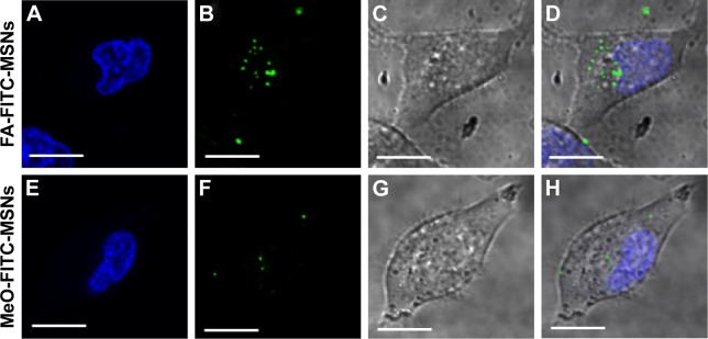
Figure 5.
Localization of FA-FITC-MSNs or MeO-FITC-MSNs in HeLa cancer cells.
Notes: Cells were exposed to 100 μg/mL of MSN materials for 6 hours. (A, E) Nucleus (blue); (B, F) FITC-MSNs (green); (C, G) DIC; (D, H) merged. Scale bars represent 5 μm.
Abbreviations: FA, folic acid; FITC, fluorescein isothiocyanate; MSNs, mesoporous silica nanoparticles; MeO, methoxy; DIC, differential interference contrast.

