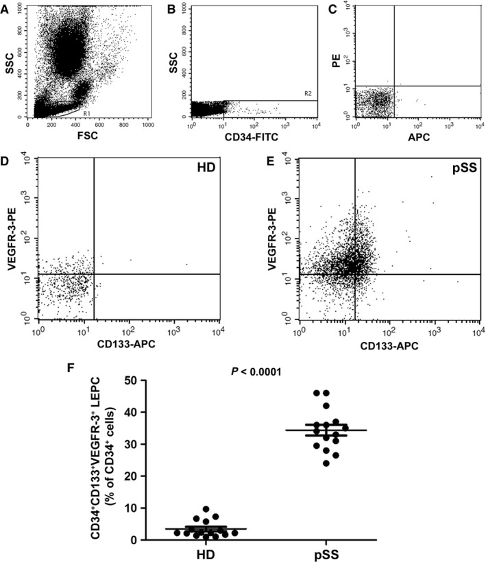
Figure 1.

Evaluation of lymphatic endothelial precursor cells (LEPCs) in the peripheral blood of patients with primary Sjögren's syndrome (pSS) and healthy donors (HD). CD34+ CD133+ VEGFR‐3+ LEPCs were identified by flow cytometry within the lymphocyte gate (A). (B) Representative flow cytometry dot plot displaying the gate drawn to select total CD34+ cells among which CD133+ VEGFR‐3+ cells (LEPCs) were identified. (C) Isotype controls. (D and E) Representative flow cytometry dot plots of one HD (D) and one patient with pSS (E). (F) CD34+ CD133+ VEGFR‐3+ LEPCs (expressed as % of total CD34+ cells) are significantly increased in the peripheral blood of patients with pSS compared with HD. Data are shown as dot plots with mean ± S.E.M. Each dot represents a participant. Mann–Whitney U‐test was used for statistical analysis.
