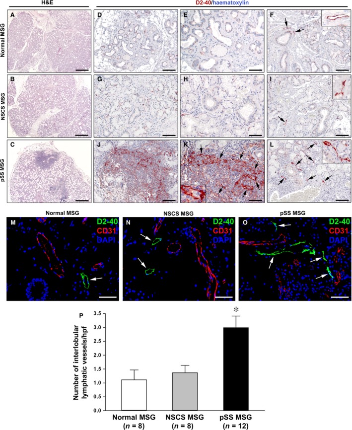
Figure 4.

Histopathology and characterization of lymphatic vascularization of minor salivary glands (MSGs). (A, D, E and F) Normal MSGs. (B, G, H and I) MSGs from non‐specific chronic sialadenitis (NSCS). (C, J, K and L) MSGs from primary Sjögren's syndrome (pSS). (A–C) Hematoxylin and eosin staining. pSS MSGs display periductal inflammatory aggregates (foci) replacing the secretory units. (D–L) Immunoperoxidase staining for the lymphatic vessel marker podoplanin (D2‐40; brownish‐red) with hematoxylin counterstain. In normal and NSCS MSGs, lymphatic vessels are absent from acinar regions (D, E, G and H), while a few lymphatic vessels (arrows) are present in the interlobular connective tissue, especially around excretory ducts (F and I). The insets show higher magnification views of lymphatic vessels from the corresponding panels. In pSS MSGs, a newly formed lymphatic capillary network (arrows) is present within periductal inflammatory infiltrates (J and K). One lymphatic capillary from the corresponding panel is shown at higher magnification in the inset; note the presence of some lymphocytes within the lumen. Numerous lymphatic vessels (arrows) are observed in the interlobular connective tissue of pSS MSGs (L). The inset shows a higher magnification view of a periductal lymphatic vessel from the corresponding panel. (M–O) Double immunofluorescence staining for D2‐40 (green) and CD31 (red) with 4′,6‐diamidino‐2‐phenylindole (DAPI, blue) counterstain for nuclei. MSG lymphatic vessels are strongly immunostained by D2‐40 (arrows), while CD31+ blood vessels are negative for D2‐40. Original magnification: ×5 (A–C), ×20 (D, F, G, I, J and L), ×40 (E, H, K and M–O), ×63 (F, I, K and L insets). Scale bar: 400 μm (A–C), 100 μm (D, F, G, I, J and L), 50 μm (E, H, K and M–O). (P) Quantification of interlobular lymphatic vessels in normal (n = 8), NSCS (n = 8) and pSS (n = 12) MSGs. Data are mean ± S.E.M. of D2‐40‐positive lymphatic vessel counts per high‐power field (hpf). *P < 0.01 versus normal and NSCS MSGs (Mann–Whitney U‐test).
