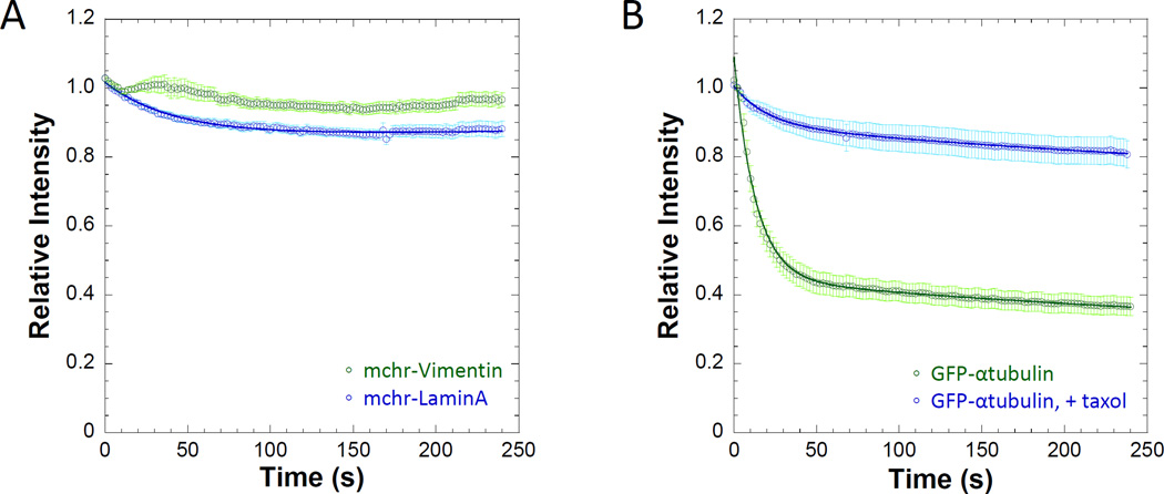
Figure 4. Cytoskeletal structures containing fluorescently labeled subunits continue to persist following 25µM digitonin permeabilization.
A) Nuclear (mchr-lamin A) and cytosolic (mchr-vimentin) intermediate filaments are both display a large insoluble fraction following permeabilization, indicating extremely slow exchange with soluble pools. B) The dissociation kinetics of GFP-labeled α-tubulin were slowed by stabilization with 10 µM taxol, where relative intensity levels never reach 50% of initial intensity due to a large immobile fraction (0.75±0.02) as a result of stabilization. Data points represent the sample mean ± SEM. For A), nlamin= 38 cells and nvimentin= 42 cells. For B), ntubulin=22 cells and n+taxol= 26 cells.

