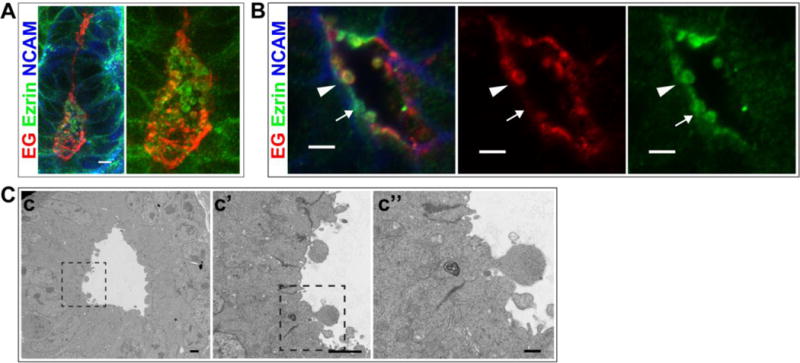
Fig 3. Endoglycan and Ezrin are present in intraluminal vesicles.

(A,B) Immunolocalization of EG and Ezrin in the intraluminal space of P0 kidney. NCAM marks the lateral plasma membrane in developing nephrons. B shows a high magnification image of a lumen (arrowhead indicates an extracellular vesicle with EG and Ezrin; arrow indicates an extracellular vesicle with Ezrin alone). (C) Transmission EM images showing intraluminal protrusions that appear to be budding from the plasma membrane. Results are representative of 2 experiments. Scale bars: 5 μm A,B; 2 μm c,c′ and 0.5 μm c″.
