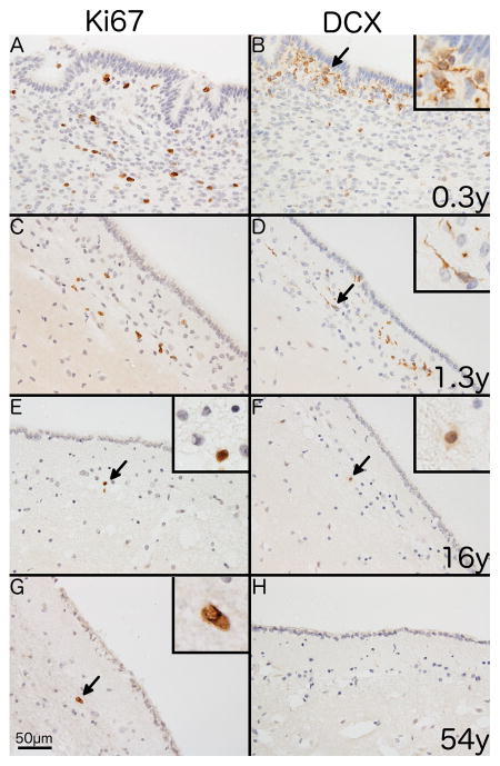
Fig. 1. Immunostaining of endogenous markers of proliferation and neurogenesis in the human SVZ.
Representative photomicrographs showing differences in immunostaining for Ki67 (A, C, E and G) and DCX (B, D, F and H) with age. Immunostaining of the SVZ from a 0.3 year-old individual shows (A) numerous Ki67+ cells with a nuclear staining pattern and (B) clusters of DCX+ cells with a combination of somatic and dendritic staining. A similar pattern is shown in a 1.3 year-old individual for both Ki67 (C) and DCX (D). A 16 year-old individual shows (E) a single Ki67+ cell (black arrow) in the SVZ and (F) shows a single DCX+ cell (black arrow) with weak somatic staining. A 54 year-old individual shows (G) a single Ki67+ cell (black arrow) and (H) no DCX+ cells. 400x magnification. Scale bar = 50μm. Insets show digital enlargement of immunopositive cells.

