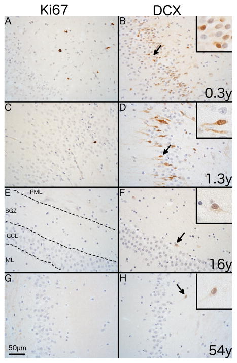
Fig. 4. Immunostaining of endogenous markers of proliferation and neurogenesis in the human SGZ.
Representative photomicrographs showing differences in immunostaining for Ki67 and DCX with age with the polymorphic layer to the right side in all images. Immunostaining shows the SGZ of a 0.3 year-old individual with (A) numerous Ki67+ cells with a nuclear staining pattern and (B) abundant DCX+ cells with predominantly somatic staining. A 1.3 year-old shows (C) a single Ki67+ cell and (D) numerous DCX+ cells with predominantly cytoplasmic staining. A16 year-old individual shows no Ki67+ cells (E) and a single DCX+ cell with dendritic and somatic staining (arrow). A 54 year-old individual shows the same pattern for both Ki67 (G) and DCX (H) as the 16 year-old individual. 400x magnification. Scale bar = 50μm.

