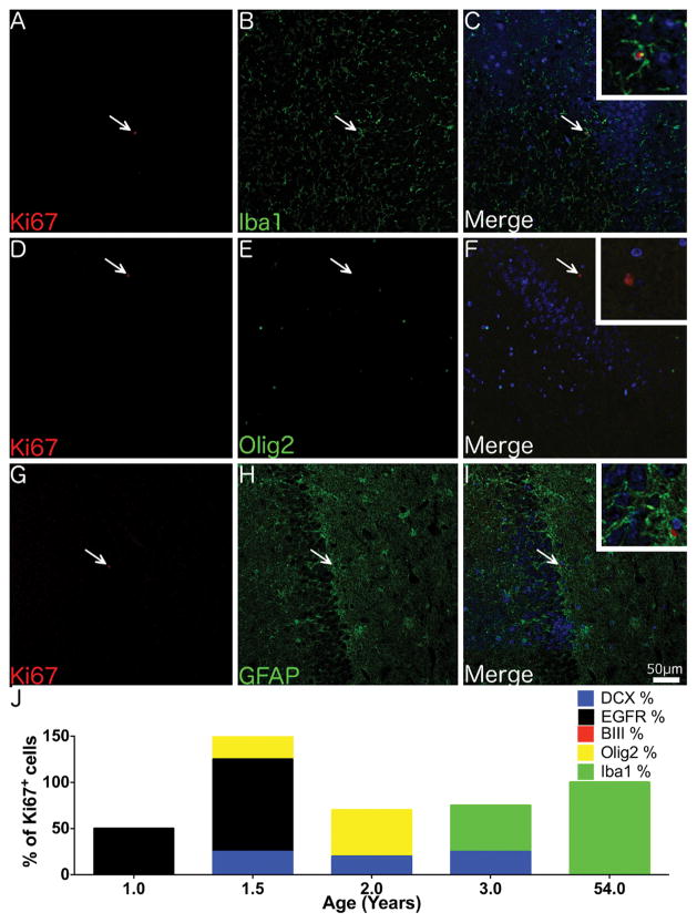
Fig. 6. Phenotype of proliferating cells in the adult SGZ.
Confocal micrographs of the SGZ from a 54 year-old individual shows Ki67+ cells co-localising with the microglial marker Iba1 (A–C) but not the immature neuronal marker beta III tubulin (D–F) or the astrocytic marker GFAP (G–I). (J) A graph displaying the number of Ki67+ cells co-localising with different cell specific markers in the SGZ across a range of ages. N.B. as quantification was performed on serial tissue sections totals could exceed or be less than 100%. (A–I) Magnification = 200x. Insets at 600x magnification. Scale bar = 50 μm.

