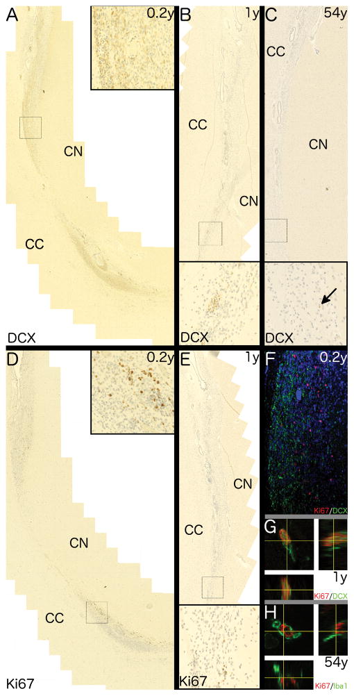
Fig. 7. Characterisation of the RMS.
Photomicrographs show traces of the rostral migratory stream with distinctive ependymal islets extending rostrally from and confluent with the frontal horn of the lateral ventricle between the corpus callosum (CC) and head of the caudate nucleus (CN). Higher magnifications of regions corresponding to black rectangles are shown in insets. Collages of overlapping fields show DCX+ cells throughout the dorsomedial portion of the RMS in a (A) 0.2 year-old and (B) one year-old brain but only a single DCX+ cell (nuclear) in the (C) 54 year-old. (D – E) Ki67+ cells are seen in ventrolateral portion of the RMS in (D) 0.2 year-old but markedly fewer Ki67+ cells within the RMS of (E) a one year-old. (F) a confocal micrograph showing the disparity of Ki67 (red) and DCX (green) within the RMS of a 0.2 year-old individual. Orthogonal projections of z-stacks show (G) a rare Ki67+/DCX+ cells in the infant RMS whilst and (H) a Ki67+ cellsin the adult RMS co-positive for the microglia marker, Iba1.

