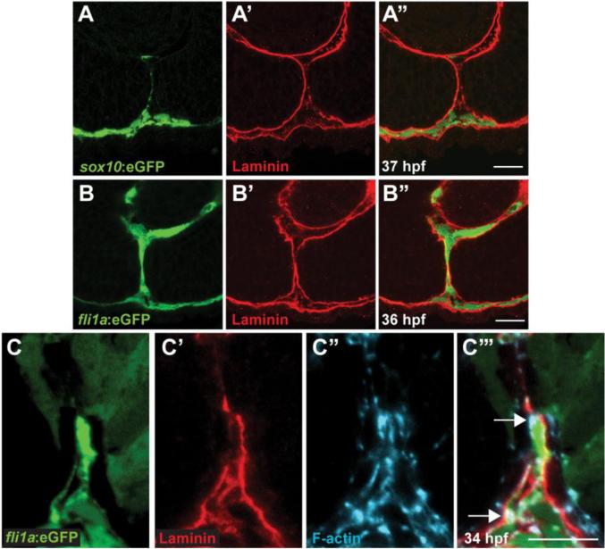
Figure 4. Periocular mesenchymal cells contribute to CFC.
(A-C) Sagittal views of the CF stained with anti-GFP (green), Lam-111 (red) and/or phalloidin (blue). (A) Few sox10:eGFP+ cells are detected in the CF, 37hpf section pictured. (B) fli1a:eGFP+ cells are retained in the CF. 36hpf section pictured. (C) fli1a:eGFP+ cells possess F-actin accumulations that localize to regions of BM breakdown. Arrows denote puncta of F-actin where Lam-111 is low or absent. 34hpf section pictured. Scale bar = 20μm (A,B) and 10μm (C).

