Fig. 4.

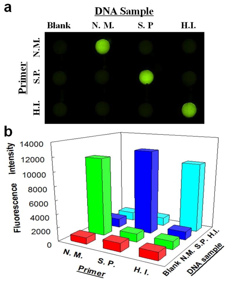
Specificity investigation among N. meningitidis, S. pneumoniae and Hib with their corresponding and non-corresponding LAMP primers by a fluorescence image (a) and fluorescence intensities (b). Lateral rows from the top to the bottom were pre-loaded with LAMP primers for N. meningitidis (N.M.), S. pneumoniae (S.P.) and Hib (H.I.) respectively; columns from left to right were introduced with Blank, DNA samples of N. meningitidis (N.M.), S. pneumoniae (S.P.) and Hib (H.I.), respectively. The RSDs for N. meningitidis, S. pneumoniae and Hib are 3.6%, 3.8% and 12.0% (n=3) on a different microfluidic device designed for the specificity test (see Fig. S4).
