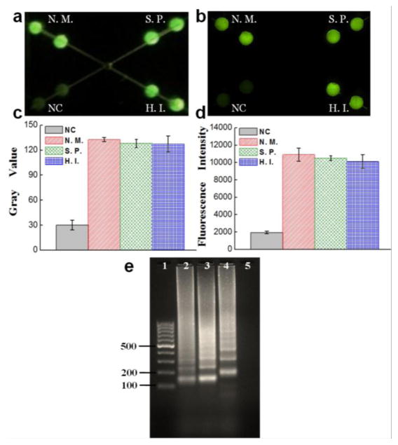
Fig. 5.
Multiplexed detection of microorganisms of N. meningitidis (N.M.), S. pneumoniae (S.P.) and Hib (H.I.) spiked in ACSF samples by a smartphone camera (a) and a fluorescence microscopy (b). (c) Gray values of different LAMP zones measured by ImageJ from a photograph taken by a smartphone camera. (d) Fluorescent intensities of different LAMP zones measured by a fluorescence microscope. (e) Gel electrophoresis analysis of LAMP products from N. meningitidis, S. pneumoniae and Hib in ACSF. Lanes 1–5: 100 bp ladder, LAMP products of N. meningitidis, S. pneumoniae and Hib in ACSF, NC.

