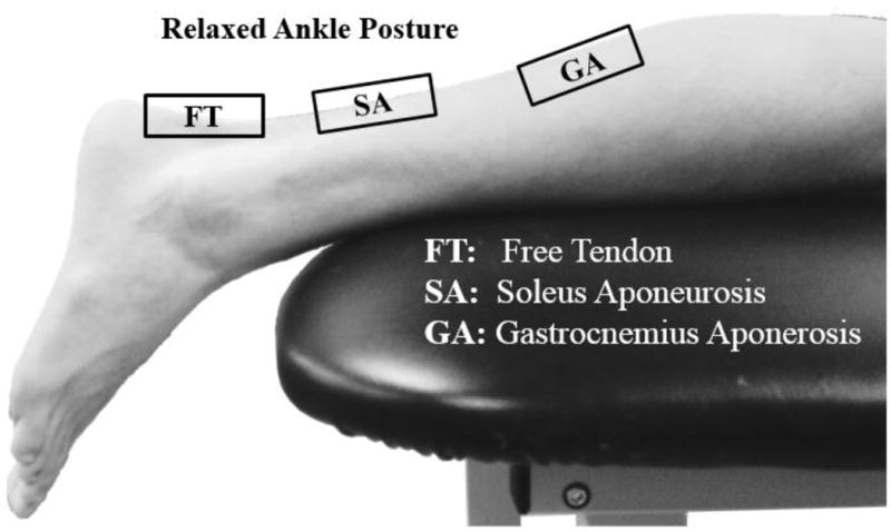
Figure 1.

Subjects were asked to lie prone on an examination table. The resting ankle posture is shown here, in which the foot is extended off the edge of the table. Shear wave data were collected from the free tendon, soleus aponeurosis and medial gastrocnemius aponeurosis.
