
Figure 1.
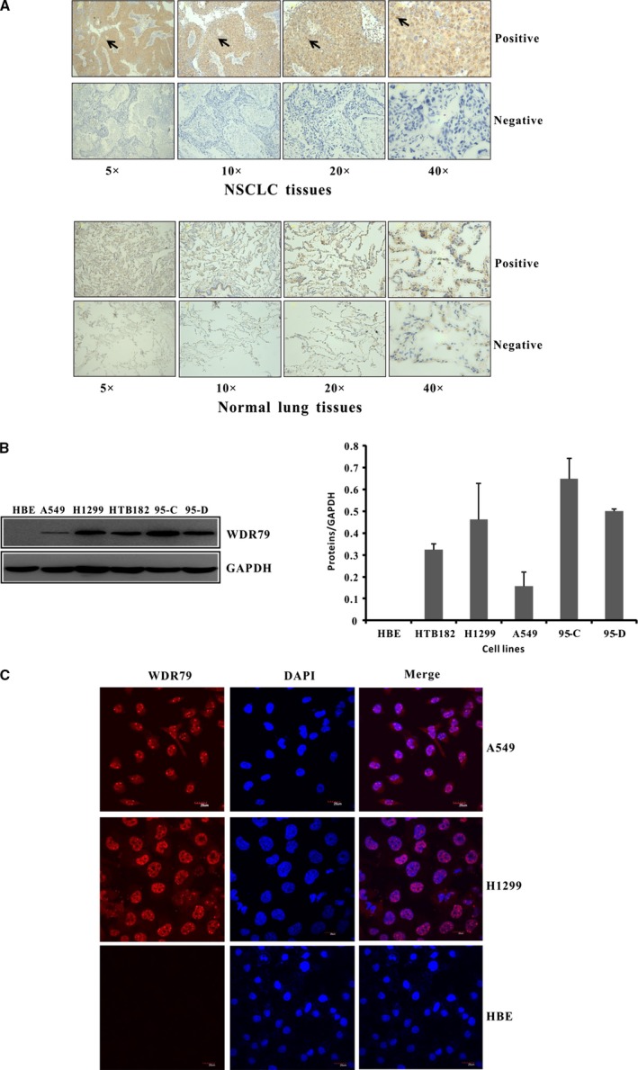
The expression and localization of WDR79 protein in NSCLC. (A) Representative IHC staining results for WDR79 in human NSCLC tissues and corresponding normal lung tissues are shown. (B) The expression level of WDR79 protein in HBE, HT182, H1299, A549, 95‐C and 95‐D cells was examined by Western blotting using WDR79 antibody. (C) A549 and H1299 cells were fixed and incubated with WDR79 antibody, followed by staining with DyLight594‐conjugated IgG for WDR79 and DAPI staining for nuclei identification.