
Figure 5.
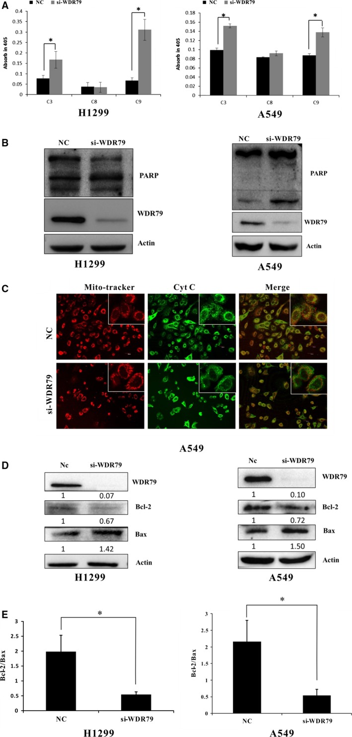
The mitochondrial pathway is involved in WDR79‐mediated apoptosis. (A) After A549 and H1299 cells were transfected with control siRNA or WDR79 siRNA for 72 hrs, caspase‐3, caspase‐8 and caspase‐9 activities were detected and presented as fold‐change relative to that of control cells, *P < 0.05. (B) A549 and H1299 cells were transfected with control siRNA or WDR79 siRNA. The expression levels of PARP were examined by Western blot using the indicated antibodies. (C) After A549 cells were transfected with WDR79 siRNA or control siRNA, cells were fixed and then incubated with cytochrome c antibody, followed by staining with DyLight488‐conjugated IgG for cytochrome c and Mitotracker for mitochondria. (D and E) After Cells were transfected with WDR79 siRNA or control siRNA, total cellular protein extracts were prepared, and the protein expression of Bcl‐2 and Bax was analysed via Western blot. Actin was used as an internal control (D). The Bax/Bcl‐2 expression ratio was quantified via densitometry (E), *P < 0.05.