
Figure 3.
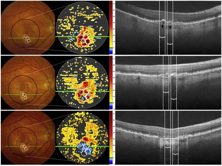
Color photographs, spectral-domain optical coherence tomography (SD OCT)—derived retinal pigment epithelium-drusen complex (RPEDC) thickness maps, and SD OCT B-scans for baseline (top row), year 2 (middle row), and year 3 (bottom row) for a single study eye. The markings follow the same designations as for Figure 1. Seven optical coherence tomography—reflective drusen substructures (ODS)-centered regions of interest (ROIs) are shown on the color photograph and SD OCT—derived thickness map at baseline (white circles, top row) and project to the white lines on the B-scan. The white circles in the middle and bottom rows track the locations of these ROIs (not new ODS) at years 2 and 3. The ODS locations at baseline are amidst drusen and some pigment clumping in this eye, without a unique characteristic on color photograph. Two low-reflective cores (ODS subtype, black arrows) are visible on the B-scan at baseline. Note that baseline positions of these ODS on the thickness maps and B-scans rapidly progress to drusen collapse at year 2 and geographic atrophy (GA) at year 3.