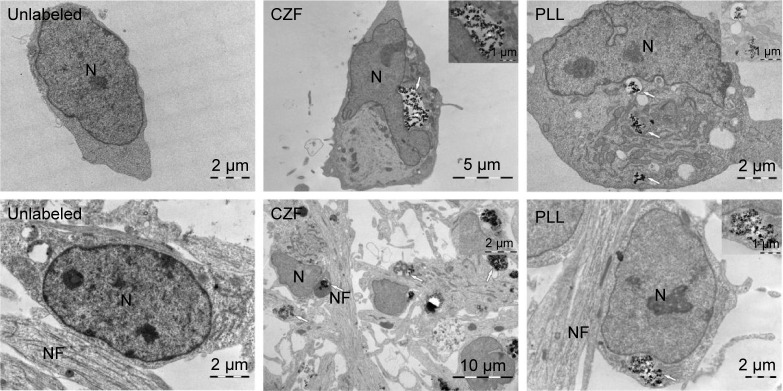
Figure 2.
TEM confirmation of nanoparticles internalization.
Notes: Top row: undifferentiated cells unlabeled and labeled with 15 μg Fe/mL CZF and PLL-coated γ-Fe2O3 (PLL) for 72 hours. Bottom row: differentiating cells unlabeled and labeled with 15 μg Fe/mL CZF and PLL-coated γ-Fe2O3 (PLL) for 72 hours. Insets show higher magnification views of nanoparticle clusters surrounded by membrane. Nanoparticle clusters are marked by arrows.
Abbreviations: TEM, transmission electron microscopy; CZF, silica-coated cobalt zinc ferrite nanoparticles; PLL-coated γ-Fe2O3, poly-l-lysine-coated iron oxide super-paramagnetic nanoparticles; N, nucleus; NF, neurofilaments.

