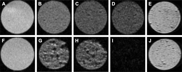
Figure 3.
MR images of gel phantoms.
Notes: Gel phantoms without cells (A) and with unlabeled cells (F). Gel phantoms with iPSC-NPs labeled with different concentrations of CZF in culture medium for 72 hours: 5 μg Fe/mL (B); 10 μg Fe/mL (C); 15 μg Fe/mL (D) and 1 week after onset of differentiation (E). Gel phantoms with iPSC-NPs labeled with PLL-coated γ-Fe2O3 at concentrations of 5 μg Fe/mL (G); 10 μg Fe/mL (H); 15 μg Fe/mL (I); and 1 week after onset of differentiation (J). Signal decrease and hypointense spots in phantoms correspond to the amount of metallic ions in cells.
Abbreviations: MR, magnetic resonance; iPSC-NPs, induced pluripotent stem cells-derived neural precursors; CZF, silica-coated cobalt zinc ferrite nanoparticles; PLL-coated γ-Fe2O3, poly-l-lysine-coated iron oxide superparamagnetic nanoparticles.

