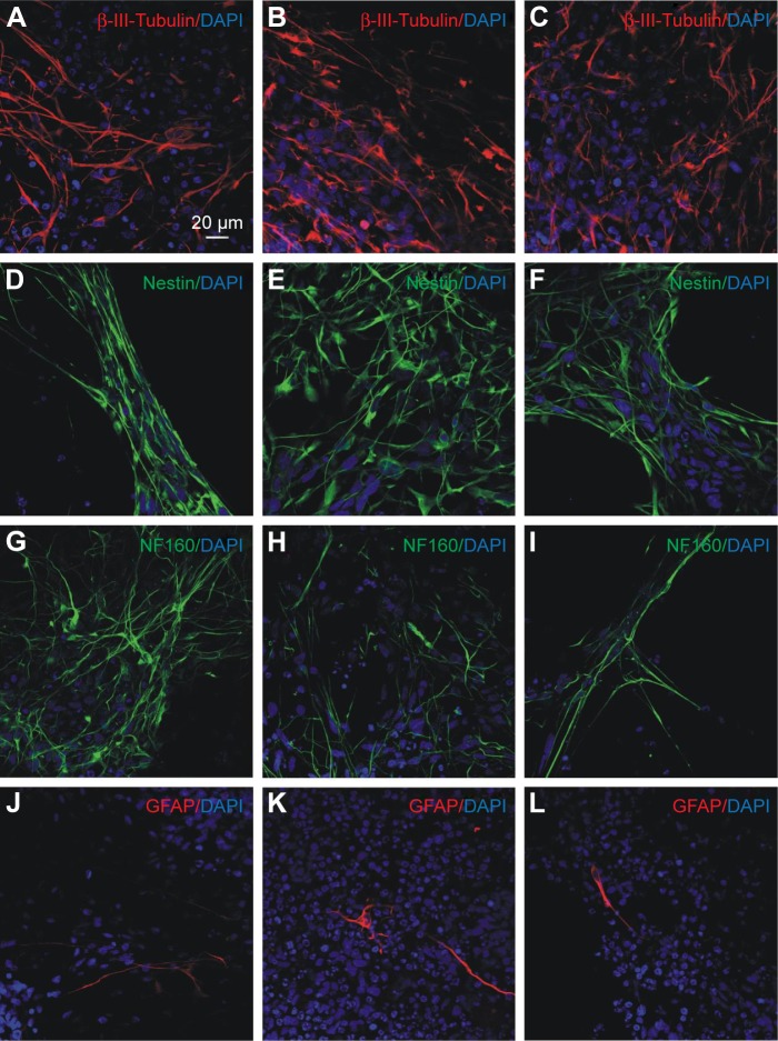
Figure 5.
Immunocytochemical characterization of differentiating iPSC-NPs.
Notes: First column (A, D, G and J) represents unlabeled cells, second column (B, E, H and K) represents cells labeled with CZF (15 μg Fe/mL in cultivation media for 72 hours), and third column (C, F, I and L) represents cells labeled with PLL-coated γ-Fe2O3 (15 μg Fe/mL in cultivation media for 72 hours). Cells are stained for β-III-tubulin (A–C) red, nestin (D–F), NF160 (G–I) green, GFAP (J–L) red and DAPI (blue) 2 weeks after onset of differentiation.
Abbreviations: iPSC-NPs, induced pluripotent stem cell-derived neural precursors; CZF, silica-coated cobalt zinc ferrite nanoparticles; PLL-coated γ-Fe2O3, poly-l-lysine-coated iron oxide superparamagnetic nanoparticles; NF160, neurofilament 160 kDa; GFAP, glial fibrillary acidic protein; DAPI, 4′,6-diamidino-2-phenylindole, dihydrochloride.

