
Figure 4.
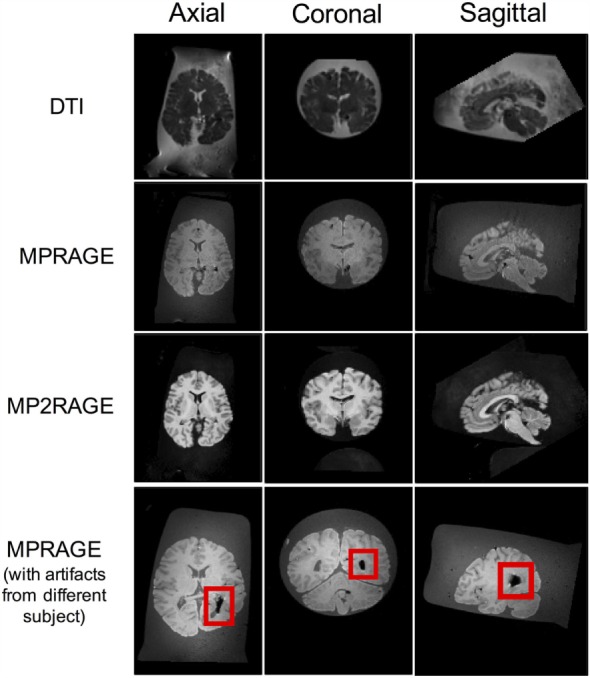
Axial, coronal, and sagittal slice views of a 71-year-old female brain acquired using the aforementioned MRI sequences following the protocols of this report. The image artifact (shown in red box) was caused by air bubbles inside the ventricle.