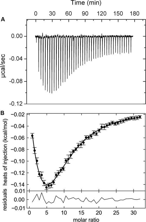
FIGURE 2.

Isothermal titration calorimetry data for the binding of αKG to the ligand binding domain of the McpK chemoreceptor. (A) Heat changes caused by the injection of 3 mM αKG into 20 μM McpK-LBD. (B) Dilution heat-corrected and concentration-normalized integrated peak areas of raw data. The solid line shows the best fit with “the two symmetric-site binding model” of the SEDPHAT program. The residual of the curve fit are shown in the lower part of the figure.
