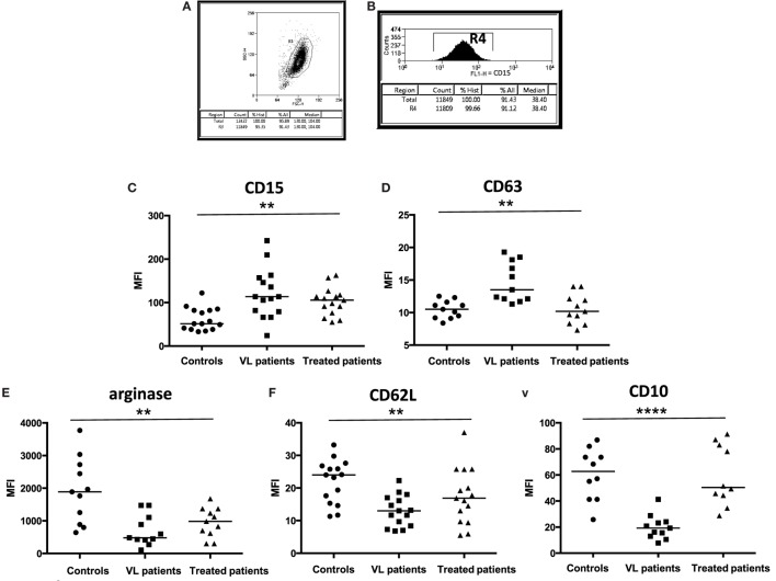
Figure 1.
Activation status of neutrophils. (A) Neutrophils were isolated by dextran sulfate sedimentation from the blood of controls, VL, and treated patients (=VL patients after successful treatment, with initial clinical cure) and (B) the frequencies of CD15+ cells were determined by flow cytometry. The expression levels (MFI = Median Fluorescence Intensity) of CD15 [(C), 15 individuals in each group], CD63 [(D), 11 individuals in each group], arginase [(E), 11 individuals in each group], CD62L [(F), 15 individuals in each group], and CD10 [(G), 10 controls, 11 VL, and 10 treated patients] were determined in the cells from gate R4 by flow cytometry. Statistical differences were determined using a Kruskal–Wallis test.

