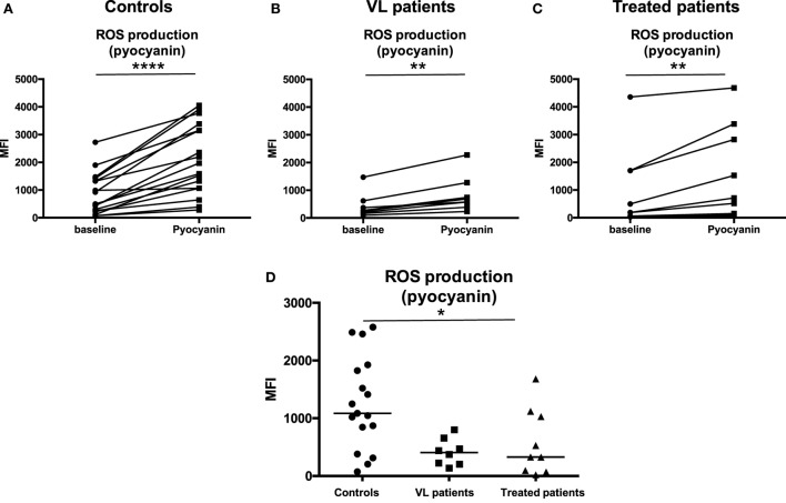
Figure 6.
ROS production in response to pyocyanin. Neutrophils were isolated by dextran sulfate sedimentation from the blood of 17 controls (A), eight VL (B), and nine treated patients [=VL patients after successful treatment, with initial clinical cure, (C)] and were incubated in the absence (baseline) or in the presence of pyocyanin. The production of ROS was evaluated by measuring the fluorescence (MFI = Median Fluorescence Intensity) resulting from the reaction of ROS with the Oxidative Stress Detection buffer. (D) MFI values were obtained by subtracting the values of obtained from the neutrophils (defined as CD15+ cells, Figure 1B) incubated without pyocyanin (baseline) from those incubated in the presence of pyocyanin. Statistical differences were determined using a Wilcoxon (A–C) or a Kruskal–Wallis test (D).

